

Knee
March 2026
Author: R. Conner Dixon, MD, CAQ-SM
Peer-Reviewer and Final Editor: Alex Tomesch, MD, CAQ-SM
Peer-Reviewer and Final Editor: Alex Tomesch, MD, CAQ-SM
This image was captured by placing a linear probe in a cranial-caudal plane in the anterior aspect of the knee below the patella.
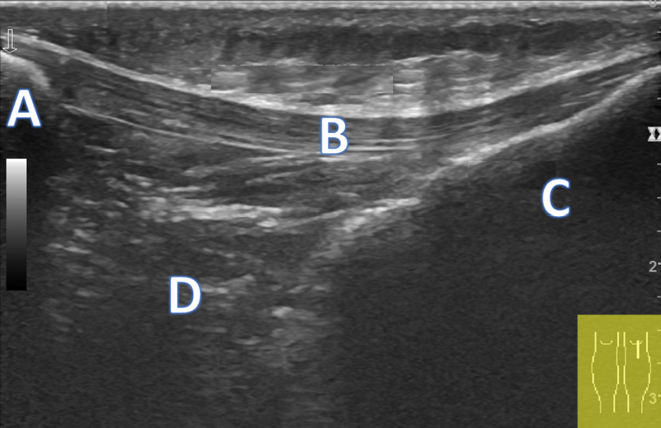
Image 1. Case courtesy of Maulik S Patel, Radiopaedia.org, rID: 86122, author’s own annotations
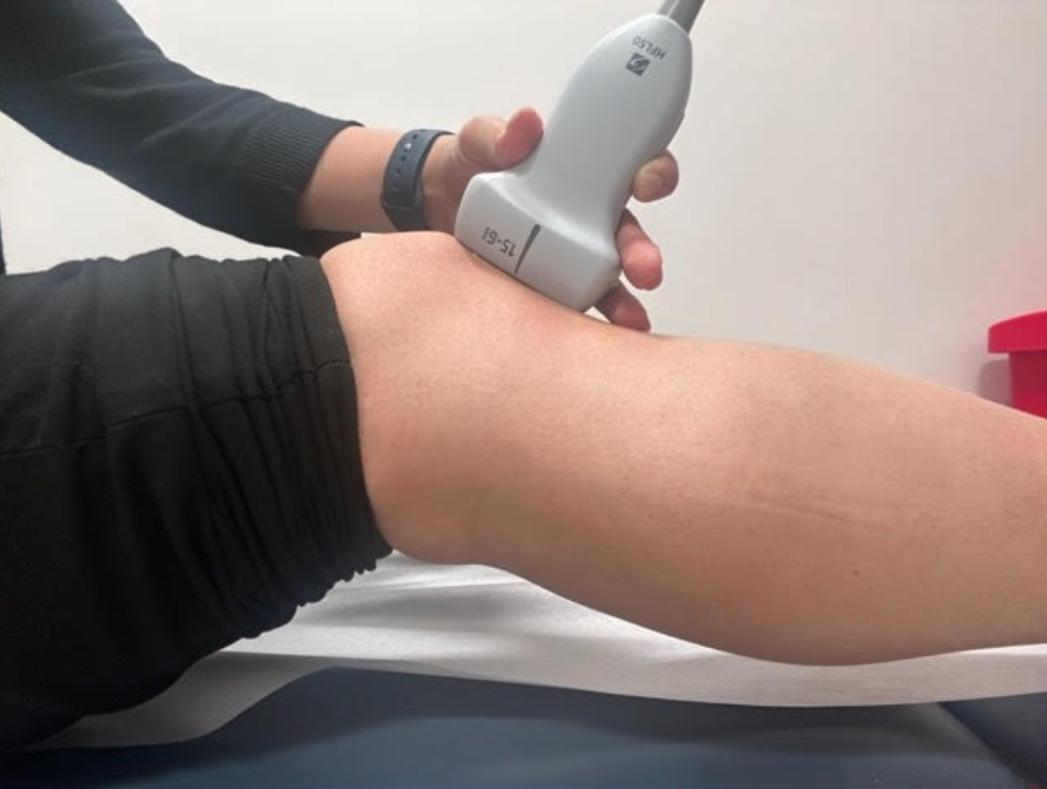
Image 2: Demonstration of the ultrasound technique for this case. Author's own image.
Can you name the structures in this longitudinal inferior knee ultrasound image?
This image was captured by placing a linear probe in a cranial-caudal plane in the anterior aspect of the knee below the patella.
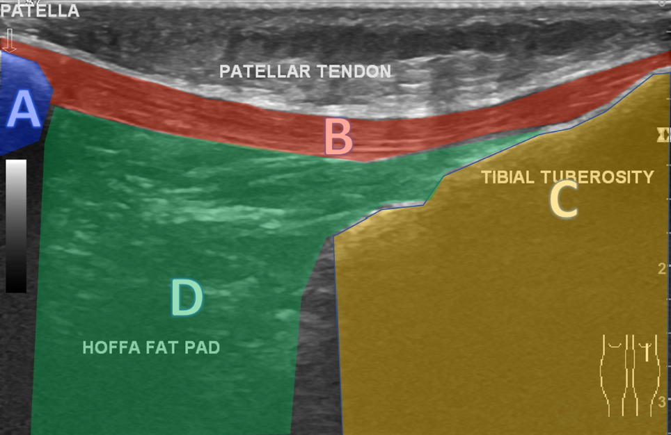
Image 3. Case courtesy of Maulik S Patel, Radiopaedia.org, rID: 86122 (author’s own annotations)
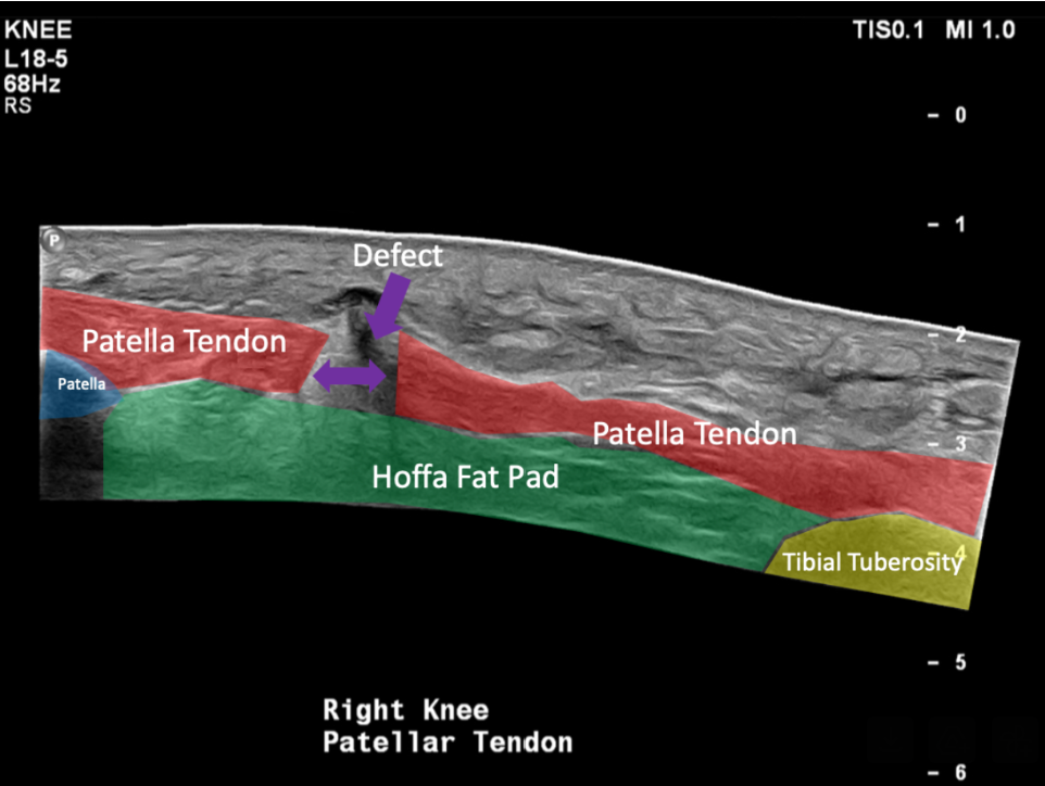
A patient comes in complaining of knee pain after stepping off a rock while hiking. She felt a pop and immediately fell, unable to put any weight on that leg. You obtain the following ultrasound image.
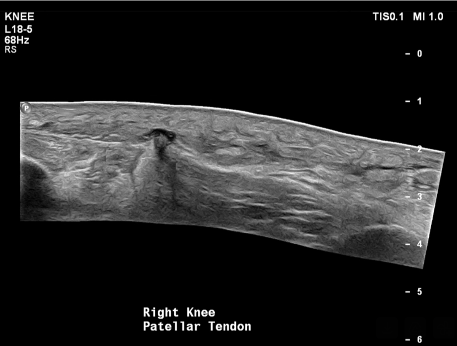
Image 4: Case courtesy of Craig Hacking, Radiopaedia.org, rID: 64416