

Use the Fascia
Author: Mark Hopkins, MD
Peer-Reviewer & Final Editor: Alex Tomesch, MD, CAQ-SM
A 91 year old female presents with right hip pain after a fall. She denies head strike and is hemodynamically stable with pulses and sensation intact.
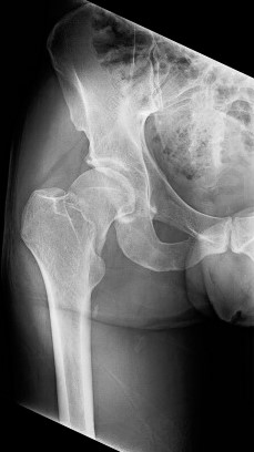
Image 1: Courtesy of Ortho EM Pearls Image Bank

What adjunct pain modalities can you use?
In contrast to use of typical pain medication, such as opioids or NSAIDs, nerve blocks have the benefit of fewer systemic effects, less need for repeat dosing, and overall greater patient pain reduction [1]. The fascia iliaca block is one of many that can help with pain management in hip fractures and can be easily performed with basic training. One study found that ultrasound guided injections by emergency physicians showed a 76% mean reduction of pain score at 120 minutes [2].
To perform this procedure you will need:
-Ultrasound machine with linear probe (may need curvilinear for larger patients)
-21 or 22 gauge spinal needle (smaller gauges may not be visible on ultrasound)
-20cc syringe
-Sterile gloves/towels/probe cover with skin cleanser
-Longer acting anesthetic (Author preference: 20cc 0.5% bupivacaine)
-Optional: 1% Lidocaine for skin surface (or can use bupivacaine)
-Adhesive bandage
Position the patient in the supine position and expose the groin on the affected side. Place the ultrasound on the opposite side of the bed to be able to view the screen in line with the procedure. Prior to sterilizing the area, find your view by first locating the femoral artery and vein as you would for a femoral central line (Image 3). The femoral nerve will be just lateral to the artery and have a “honeycomb” appearance. The fascia iliaca is the hyperechoic thin line overlying the nerve and iliopsoas muscle, then dipping posterior to the femoral artery (see Image 4 for labels).
-
Pearl: If the nerve does not first come into view, fan back and forth as an incorrect probe angle can make it difficult to visualize.

Image 3. View of right inguinal neurovascular bundle. Image by author

Image 4. FI=Fascia Iliaca. IP=Iliopsoas Muscle. FN=Femoral Nerve. AT=Adipose Tissue. FA=Femoral Artery. Image and labels by author
After verifying your location, create a sterile field and utilize the probe cover. Anesthetize the skin, then insert the needle at a 30-45 degree angle lateral to and in-plane with the probe. The needle tip should be visible on the screen at all times during the procedure. Puncture just through the fascia while staying lateral of the femoral nerve (Image 5). Inject a small amount to verify position. The anesthetic should dissect the fascial layer from the underlying iliopsoas muscle and begin spreading towards the femoral nerve. After verifying position, inject the full amount (Image 6). Withdraw the needle and place the adhesive bandage. The patient should begin to feel relief within 15-20 minutes.
-
Pearl: By staying lateral to the femoral nerve, this will also anesthetize the lateral femoral cutaneous nerve and avoid injecting into the nerve or artery.

Image 5. In plane lateral needle insertion. Image and labels by author

Image 6. Hypoechoic fluid collection after anesthetic injection. Image and labels by author
-
Pearl: If you are going to be doing these types of procedures you should be aware of symptoms of local anesthetic toxicity. Additionally, you should have intralipid readily available in the department as this is the antidote for toxicity.
References
[1] Foss NB, Kristensen BB, Bundgaard M, Bak M, Heiring C, Virkelyst C, Hougaard S, Kehlet H. Fascia iliaca compartment blockade for acute pain control in hip fracture patients: a randomized, placebo-controlled trial. Anesthesiology. 2007 Apr;106(4):773-8. doi: 10.1097/01.anes.0000264764.
[2] Haines L, Dickman E, Ayvazyan S, Pearl M, Wu S, Rosenblum D, Likourezos A. Ultrasound-guided fascia iliaca compartment block for hip fractures in the emergency department. J Emerg Med. 2012 Oct;43(4):692-7
[3] Khan SK, Kaira S, Khanna A, Thiruvengada MM, Parker MJ. Timing of surgery for hip fractures: A systematic review of 52 published studies involving 291,413 patients. Injury 2009; 40: 692–7